
Research
Publication
- Multimodal Analysis of Structural and Functional MRI for Schizophrenia Diagnosis, Huixiang Zhuang, Yao Li, Ruihao Liu, Chaowei Wu, and Manhua Liu, Tenth International Conference on Digital Image Processing (ICDIP 2018), 2018, p. 6. PDF
- Multimodal Classification of Drug-naive First-episode Schizophrenia Patients Combining Structural and Functional Magnetic Resonance Imaging, Huixiang Zhuang, Ruihao Liu, Chaowei Wu, Ziyu Meng, Danni Wang, Dengtang Liu, Manhua Liu and Yao Li, Neuroscience Letters (under review) PDF
- Alternations of Brain Structural Connectivity after Unilateral Upper-limb Amputation, Xiaoli Guo, Ruihao Liu, Jincheng Lu, Chaowei Wu, Yuanyuan Lyu, Zhuo Wang, Jianbo Xiang, Changjie Pan and Shanbao Tong, IEEE Transactions on Biomedical Engineering (TBME) (under review) PDF
Research Experience
New Strategy for Super-resolution in Magnetic Resonance Imaging, Jan 2018 - Present
In UIUC and SJTU, Advised by Prof. Zhi-Pei Liang
Keywords: MRI Image Reconstruction, Machine Learning
The proposed super-resolution pipeline is series of strategies to tackle the ill-posed problem where high-frequency component is recovered. Convolution neural network, sparsity-based model and data consistency model is combined to achieve this process. Experiment verified proposed method was able to reconstruct MPRAGE image with 4 times down-sampled k space.
- Leveraged the power of deep learning to give a very good initial estimation of the high-resolution image given the limited low-resolution image
- Extracted and recovered novel features which existed in practical patient images, based on k-space residual and sparsity limitation
- Utilized optimization model based on data consistency to eliminate artifacts coming along with high-resolution images generated by neural network and gave final prediction
More information: Presentation Slides (PDF) can be viewed here. Full Report can be viewed here.
Multi-modal Imaging Classification using Machine-learning Algorithm in Firstepisode Schizophrenia, Oct 2016 - July 2017
In SJTU, Advised by Prof. Yao Li
Keywords: Multi-modal MRI, Machine Learning, Classification, Neuroscience
Established a method to distinguish the health and first-episode schizophrenia (SZ) patients given multi-modal MRI data (fMRI, DTI, T1). Proposed multimodal classification method had 81.2% accuracy with 92.5% sensitivity and 66.7% specificity for SZ diagnosis. This project received an A (Top rank of the program). The full manuscript has been published in Tenth International Conference on Digital Image Processing (ICDIP 2018). Another manuscript is under review now.
- Exploited t-test analysis after data processing using FSL and other toolkits
- Utilized Sparse Coding algorithms to screen out potential features among various biomarkers
- Implemented Random Forest algorithms to estimate the potential relationship among selected features to reach an ideal group discriminating performance
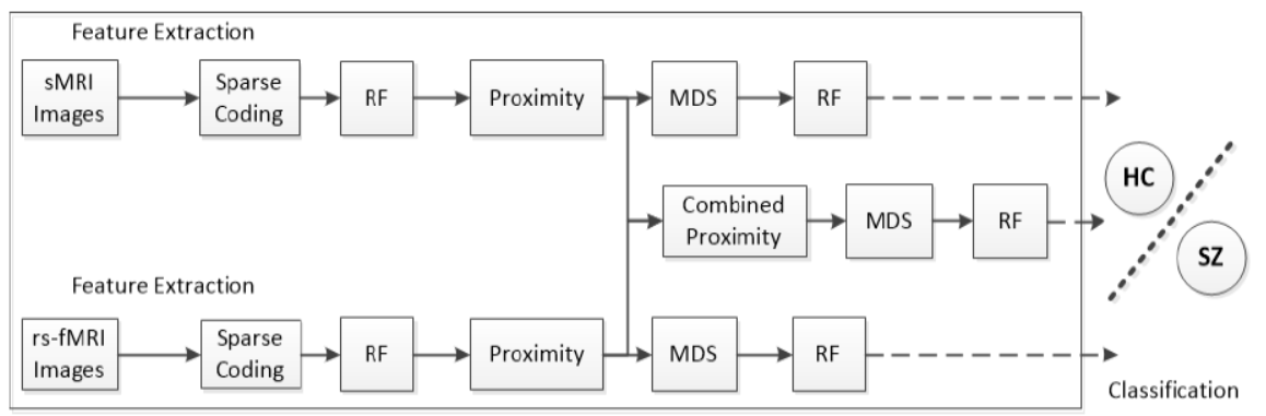
Fig.1 The framework of the proposed multi-modality classification algorithm
| Modality | Accuracy(%) | Sensitivity | Specificity | k |
|---|---|---|---|---|
| sMRI | 65.7 | 77.5 | 50.0 | 43 |
| rs-fMRI | 75.5 | 85.0 | 63.3 | 41 |
| Multi-modality | 81.2 | 92.5 | 66.7 | 48 |
Table 2 Classification performances of different modalities by the proposed method
| Modality | Without Sparse Coding | Without Multimodal Scaling | The Proposed Method |
|---|---|---|---|
| Sensitivity (%) | 87.5 | 92.5 | 92.5 |
| Specificity (%) | 51.7 | 63.3 | 66.7 |
| Accuracy (%) | 72.6 | 79.8 | 81.2 |
Table 3 Comparison of multi-modal classification accuracy among the methods
Research of Brain Microstructure Alterations for Upper-limb Amputees, July 2017 - Present
In SJTU, Advised by Prof. Xiaoli Guo
Keywords: MRI, Diffusion Tensor Imaging, Probabilistic Diffusion Tractography, Neuroscience
Analyzed white matter microstructure alterations after upper-limb amputation given DTI images. One paper in preparation; Plan to submit to journal shortly (First Author)
- Found significant lower Fractional Anisotropy (FA) and other significant different indices in subregions of corpus callosum (CC) in patients with residual limb pain
- Implemented Probabilistic Diffusion Tractography (PDT) and found similar changes in corresponding transcallosal tracts
- Indicated interhemispheric pathways contributing to pain sensation; chronic pain were reorganized in upper limb amputees
More details will be added soonly. (Last update: 1/1/2019)
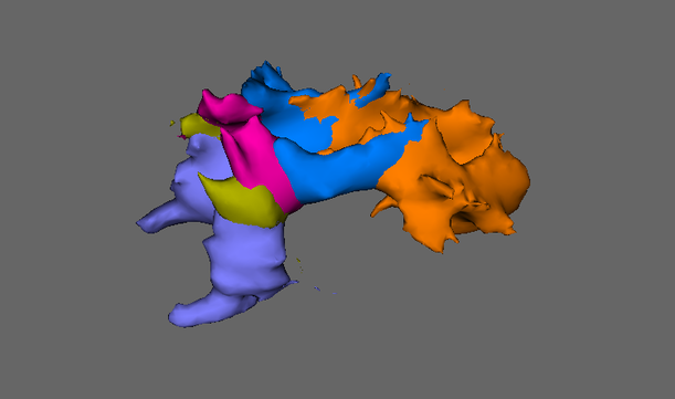
Fig.1 Transcallosal tracts connecting the unilateral area and the contralaterally homogenous area
The research and development of computer-aided orbital decompression surgery planning system
In SJTU, Advised by Prof. Lixu Gu
Keywords: Computerized Tomography (CT), Image Processing, Software Development
Developed a software specialized for measurement of intra-orbital structures. It exactly meeted clinical needs and have been applied in clinical at the Shanghai Ninth People’s Hospital.
- Realized a series functions including 3-D CT image reconstruction, coordinates correction, segment, etc. with VTK library.
- Integrated into a completely independent software with a user-friendly GUI
- Cooperated with an ophthalmologist to identify clinical needs; Effectiveness was verified with statistic analysis
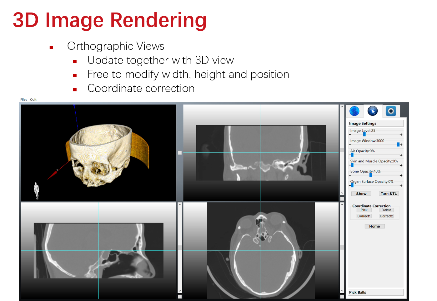
Fig.1 Function: 3D Image Rendering and features
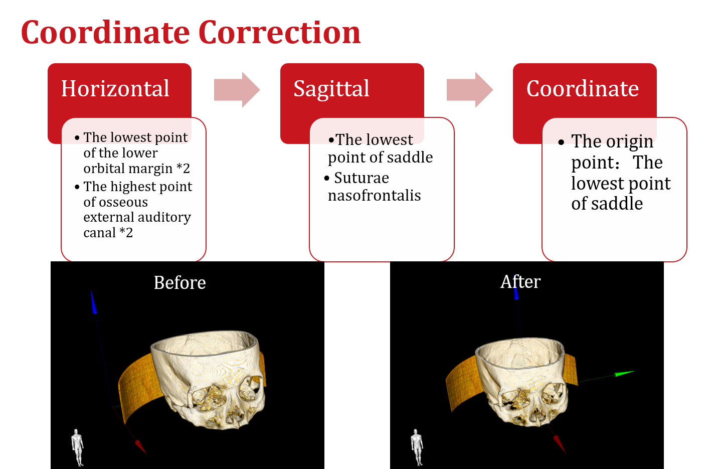
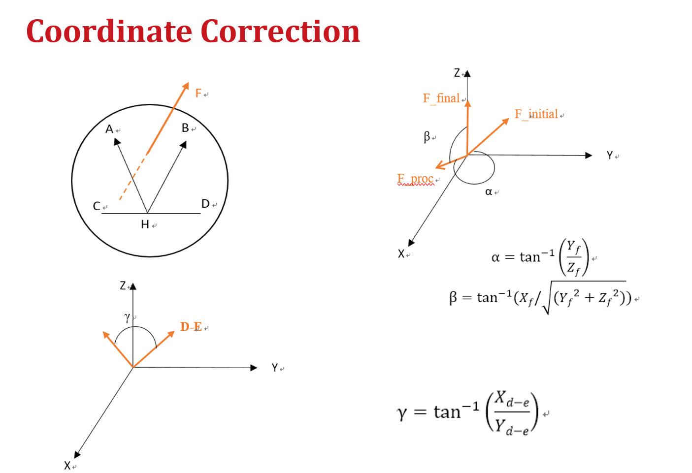
Fig.2 & 3 Function: Coordinate Correction and its principle
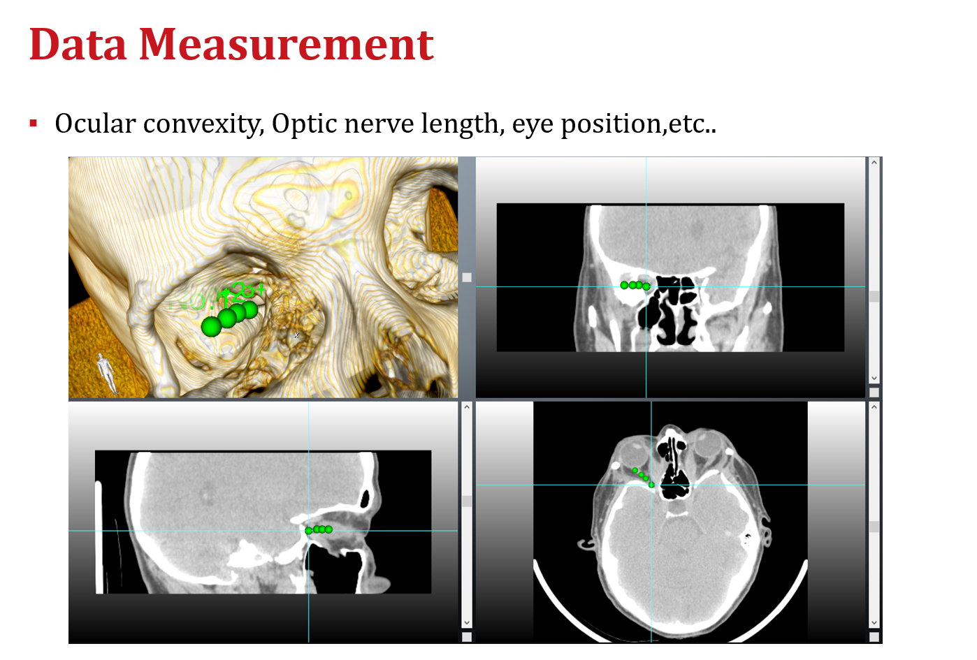
Fig.4 Function: Data Measurement
Video 1 3D Rendering
Video 2 Image Viewing and Choose Point
Video 3 Region Growth Segmentation
A universal multifactorial visualized detection system
In SJTU, Advised by Dr. Lin He and Dr. Gang Ma
Keywords: Synthetic biology, Biological Modelling, Genetic Engineering, iGEM Gold Metal
This project sought to transform genetically engineered Escherichia Coli into a visualized monitor, which could detect multiple metal ions, display the concentration as a combination of colors, and achieve quantitative measurement. While serving as the vice leader of a research team on behalf of SJTU (14 undergraduates majoring in Bioengineering, Bioinformatics, Biomedical Engineering and Material Science), I was in charge of the modeling work to explore the relationship between our aimed detection substances and chromoproteins. I orally presented our work to professional judges from all over the world and earned a Gold Medal in the International Genetically Engineered Machine (iGEM) Competition (top-level international competition in synthetic biology).
- Detected multiple metal ions at the same time, displaying the concentration as a combination of colors
- Achieved quantitative measurement by building models and developing mobile APP

Fig.1 Logo of our team
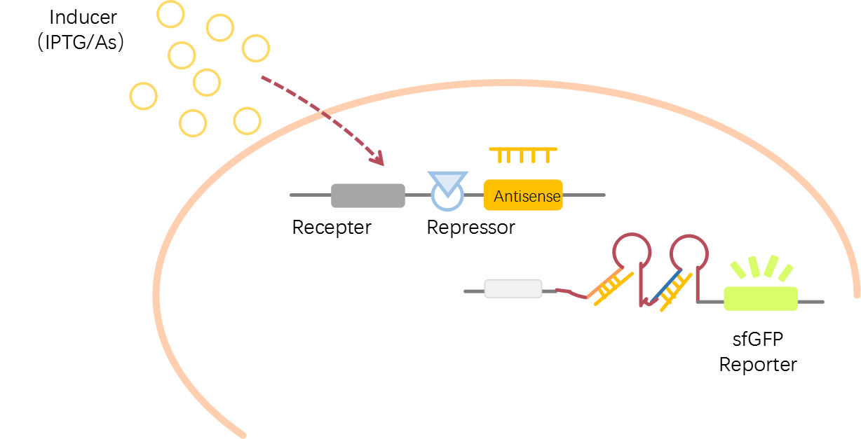
Fig.2 Signal Pathway:Inducers enters the cell, activates receptor and binding with repressor protein, leading to antisense producing. Then antisense binds with STAR. Finally sfGFP expression starts.
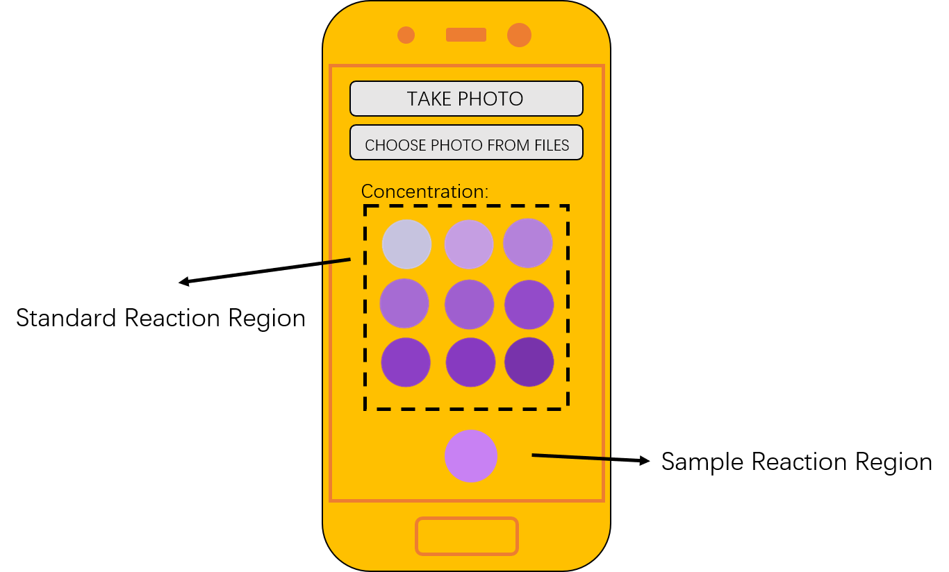
Fig.3 Diagram of developed mobile APP
More details can be viewed in Team Wiki.
Course Projects
Evaluation of the Effect of Blood Vessel Position and RF Power in Tumor Ablation Simulation
Course: Bio Heat Transfer
Keywords: Finite Element Modelling (FEM), Bioheat
- Established a RF tumor ablation model on finite element model software COMSOL Multiphysics
- Explored the effect of blood vessel position and RF power in temperature distribution and necrosis tissue fraction distribution
- Led a course group, organizing weekly group discussion and got the** highest** score of the class in project report
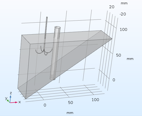
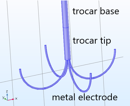
Fig.1&2 General geometry and electrode geometry of the model
Fig.3 Temperature distribution of x-z plane at 0, 1, 2, 5, 10, 20 min. (blood vessel is right of electrode). Fig.4 Necrosis tissue fraction, respectively.
Fig.5 Temperature of one electrode arm tip. Fig.6 Temperature contour in 1, 2, 5, 10 min.
Fig.7 Simulation of temperature of human in freezing water and cold air(Note: your seeing is just surface temperature)